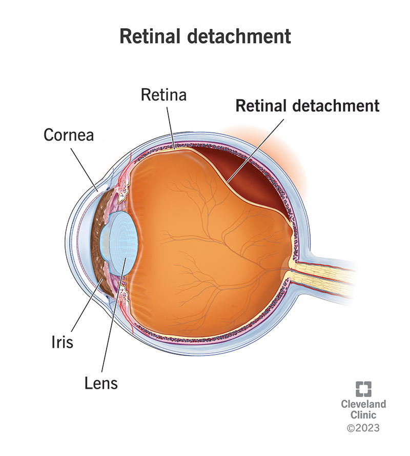
Figure 1. [The normal human retina fundus]. - Webvision - NCBI
Por um escritor misterioso
Last updated 08 novembro 2024
![Figure 1. [The normal human retina fundus]. - Webvision - NCBI](https://www.ncbi.nlm.nih.gov/books/NBK554706/bin/Archetecture_Fovea-Image006.jpg)
The normal human retina fundus photo shows the optic nerve (right), blood vessels and the position of the fovea (center).
![Figure 1. [The normal human retina fundus]. - Webvision - NCBI](https://www.mdpi.com/cells/cells-12-01987/article_deploy/html/images/cells-12-01987-g001.png)
Cells, Free Full-Text
![Figure 1. [The normal human retina fundus]. - Webvision - NCBI](https://pub.mdpi-res.com/diagnostics/diagnostics-13-02373/article_deploy/html/images/diagnostics-13-02373-g001.png?1689332399)
Diagnostics, Free Full-Text
![Figure 1. [The normal human retina fundus]. - Webvision - NCBI](https://webvision.med.utah.edu/wp-content/uploads/2018/07/Fig-14-macula-lutea.jpg)
Simple Anatomy of the Retina by Helga Kolb – Webvision
![Figure 1. [The normal human retina fundus]. - Webvision - NCBI](https://www.frontiersin.org/files/Articles/895519/fimmu-13-895519-HTML/image_m/fimmu-13-895519-g001.jpg)
Frontiers As in Real Estate, Location Matters: Cellular Expression of Complement Varies Between Macular and Peripheral Regions of the Retina and Supporting Tissues
![Figure 1. [The normal human retina fundus]. - Webvision - NCBI](https://www.researchgate.net/publication/333702798/figure/fig1/AS:771957922480128@1561060519865/The-human-retina-with-different-stages-of-NPDR-a-normal-retina-and-its-main-components.png)
The human retina with different stages of NPDR: a normal retina and its
![Figure 1. [The normal human retina fundus]. - Webvision - NCBI](https://www.ncbi.nlm.nih.gov/books/NBK554060/bin/466648_1_En_8_Fig1_HTML.jpg)
Fig. 8.1, [Cross-sectional OCT image of human retina with the corresponding cellular structures]. - High Resolution Imaging in Microscopy and Ophthalmology - NCBI Bookshelf
![Figure 1. [The normal human retina fundus]. - Webvision - NCBI](https://www.ncbi.nlm.nih.gov/books/NBK1222/bin/retinoschisis-Image001.jpg)
Figure 1. [Fundus photo of a male]. - GeneReviews® - NCBI Bookshelf
![Figure 1. [The normal human retina fundus]. - Webvision - NCBI](https://www.researchgate.net/publication/242466981/figure/fig1/AS:298496365219842@1448178487460/A-Normal-fundus-of-OD-B-Fundus-of-OS-showing-foveal-retinal-pigment-epithelial.png)
A) Normal fundus of OD; (B) Fundus of OS showing foveal retinal
![Figure 1. [The normal human retina fundus]. - Webvision - NCBI](https://www.ncbi.nlm.nih.gov/books/NBK11533/bin/sretinaf20.gif)
Simple Anatomy of the Retina - Webvision - NCBI Bookshelf
![Figure 1. [The normal human retina fundus]. - Webvision - NCBI](https://media.springernature.com/m685/springer-static/image/art%3A10.1186%2Fs12877-021-02009-z/MediaObjects/12877_2021_2009_Fig1_HTML.png)
Association of reduced retinal arteriolar tortuosity with depression in older participants from the Northern Ireland Cohort for the Longitudinal Study of Ageing, BMC Geriatrics
Recomendado para você
-
Retinal Detachment: Symptoms & Causes08 novembro 2024
-
 What Is a Detached Retina? - Outlook Eyecare08 novembro 2024
 -
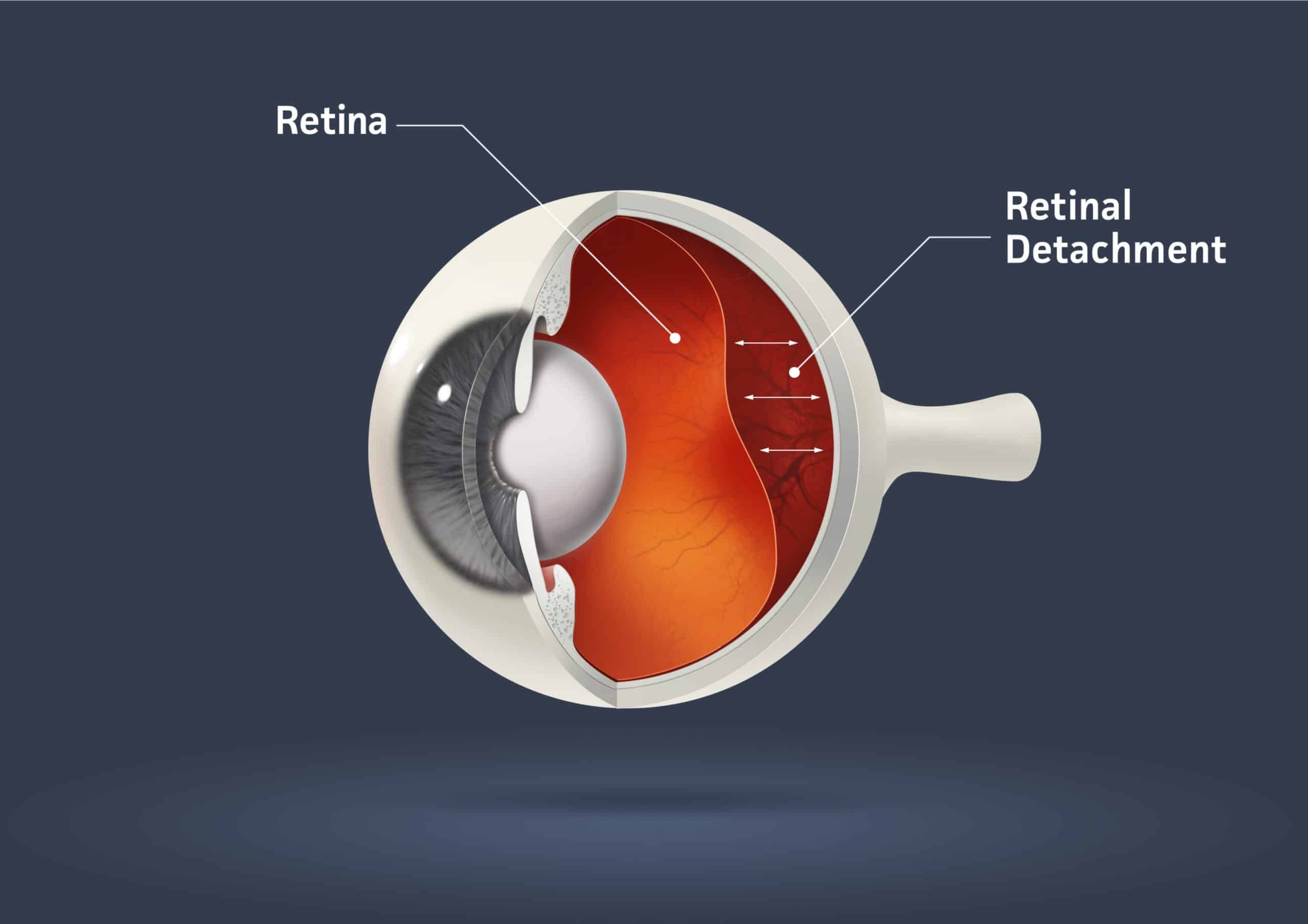 Can You Prevent and Treat Retinal Detachment?08 novembro 2024
 -
 Types of Retinal Detachment, Their Causes, and Treatments08 novembro 2024
 -
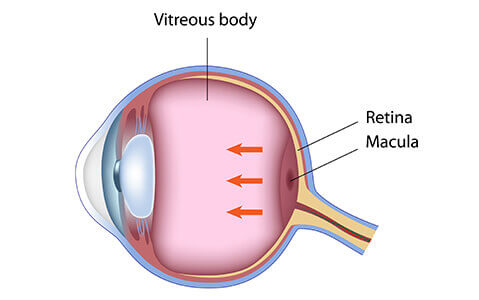 Retina Lee Eye Center08 novembro 2024
 -
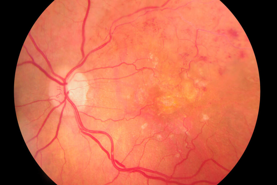 Descolamento de retina: causas, sintomas, tratamentos e recomendações08 novembro 2024
 -
 Retina Definition, Anatomy & Function - Video & Lesson08 novembro 2024
 -
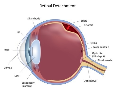 What Causes Retinal Detachment?08 novembro 2024
 -
 51,730 Retina Images, Stock Photos, 3D objects, & Vectors08 novembro 2024
 -
 Retina e Vitreo - Clínica de Olhos Nações08 novembro 2024
você pode gostar
-
 Kirby and the Forgotten Land Review – Kirby Doesn't Suck! – WGB08 novembro 2024
 -
Meaning of Cool by Dua Lipa08 novembro 2024
-
 Seattle facing rise in fentanyl-related overdoses, running out of space to store dead bodies08 novembro 2024
 -
 Ao Haru Ride Light Novel08 novembro 2024
 -
 PS4 Archives - Average Joe Games08 novembro 2024
 -
 Imagens Engraçadas As Fotos Mais Engraçadas da Internet em 202308 novembro 2024
 -
 Friday Night Funkin' HD 🔥 Jogue online08 novembro 2024
 -
YUMI. YUMI. SIGNIFICADO DO NOME YUMI Yumi: Significa “beleza08 novembro 2024
-
 Beatriz Milz - Buscando informações na Wikipédia: Lista de episódios de Naruto Shippuden08 novembro 2024
 -
 Doodle Jump Episode 208 novembro 2024
